Comprehensive Eye Exam
Comprehensive Eye Exam
Your Eye Exam Explained
Every eye examination is tailored to your individual needs.
At the beginning of the eye exam, a pre-test is usually performed. The doctor uses the results of the pre-test during the eye examination to thoroughly check the health of your eyes and give you the best possible vision correction.
Depending on your individual needs, a variety of different pieces of diagnostic equipment may be used during the pre-test including:
- OCT scanner
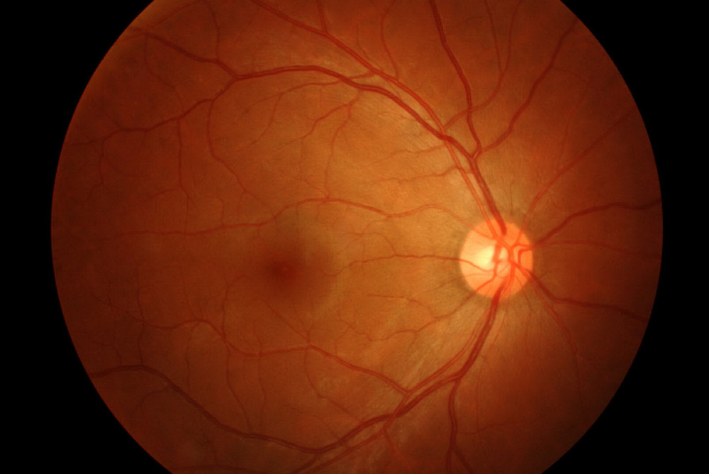
- Fundus Photographer
- Autorefractor
- Tonometer
- Pachymeter
- Perimeter
An OCT machine can take a scan of the back layers of your eyes and a photograph. These images are saved and will be useful in monitoring changes in eye health on future visits.
Autorefraction indicates how long or short-sighted someone is. Autorefractors give highly accurate measurements that are used to determine vision correction needs.
Autorefractors only take a few moments to determine each measurement for each eye and are used in conjunction with a machine called a phoroptor to switch lenses in front of your eyes to provide ideal vision correction. Tonometry allows an eye doctor to check the eye pressure which is an important test for glaucoma screening. Pachymeter checks the thickness of the front transparent layer of your eye and a perimeter checks your field of vision.
Your eye doctor will check the health of your eyes and look for signs of other medical conditions. They will examine the retina at the back of your eye, the optic nerve and its blood vessels and the front surface of the eyes.
Your eye doctor might need to apply eye drops to dilate your pupils. This can make your eyes a bit blurry and more sensitive to light, which could affect driving. The eye doctor will inform you if they are going to dilate your eyes and recommend ask a friend or family member to drive you.
At the end of your eye examination, your eye doctor will explain what all the tests and evaluations have indicated and recommend the very best options for your individual needs.
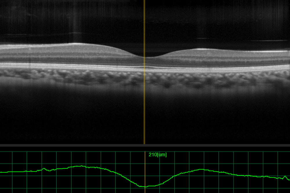
What is an OCT Scan?
Optical Coherence Tomography (OCT) is a non-invasive imaging test. OCT uses light waves to take cross-section pictures of your retina. This scan allows the eye doctor to see detailed images of the retina (the innermost layer of the interior eye), enabling us to accurately detect, monitor and control changes to the retina. OCT is also useful in detecting disorders of the optic nerve (the collection of nerve fibres which send signals from the retina to the brain).
What eye conditions can an OCT Scan help to detect?
This provides diagnosis, management, monitoring and treatment guidance for many conditions like:
- Age related macular degeneration.
- Diabetic eye disease
- Glaucoma

Eye Exam